The early use of x-rays for imaging and the treatment of cancer
Page uploaded 15th December 2013
Back to historic events in Exeter
Many would think that, in the past, the Royal Devon and Exeter Hospital, being located in the sleepy county of Devon, was not at the leading edge of research and patient care. This is not altogether true, for since 1740, when the hospital was founded, some physicians and surgeons have pushed the boundaries of research, to improve the care of their patients.
One such area of expertise, was in the provision of x-ray diagnosis and treatment. Just one year after Röntgen first investigated x-rays, the first demonstration of the new technology was given in Exeter in October 1896, to an audience at the Working Men’s Society.
The first diagnostic x-ray
The first x-ray taken at the hospital, was a 48 minute exposure of a fractured radius on 3 February 1898. An amateur photographer, Mr Benjamin Benford was asked to take the image for the surgeon Mr Russell Coombe.
In 1902, Mr Percy Stirk, a surgeon at the hospital, gave evidence at a court, regarding a work injury. He showed x-rays of an injury to the judge and jury. The defendant’s barrister remarked of the injuries shown “They are very horrible”. It is probable that Mr Stirk was using an early x-ray apparatus at his private practice, rather than at the hospital, as the new technology was closely allied to photography.
Another court case to which Mr Stirk gave evidence.
Exeter and Plymouth Gazette - 28 June 1904
“Mr. P. Stirk, house surgeon at the Royal Devon and Exeter Hospital, said that when examining the prosecutrix at the institution he found a small puncture wound in the back of the right shoulder, midway between the shoulder blade and the spine. There was some swelling and the parts were very tender. On examining the wound by means of X-Rays the witness saw a piece of metal deeply embedded in the muscles of the back, and on being extracted, was found to be a piece of pin about an inch in length. The end of the broken pin was one. and half inches below the skin, and the point was practically touching the rib. To have inserted the pin would have needed considerable force.”
The first x-ray pioneer
In July 1907, another surgeon, Dr J Delprat Harris, was reported that he “then gave some interesting experiments with the Rontgen and X Rays, after which a marble tablet, recording the event, was unveiled by Lady Duckworth King.” Dr J Delprat Harris was from 1881, an ophthalmic surgeon at the hospital, and later, a general surgeon, following his grandfather, who had also been a surgeon.
The Electrical Department, under the supervision of Delprat Harris, was opened in 1907, with funds provided by the widow of the banker Mr S A Sanders—the department also handled the responsibility of the early x-ray equipment at the hospital.
Harris continued his use of x-ray equipment, for clinical diagnosis. Still a novelty, the newspapers would often report on its use, especially after street accidents. In February 1914, Delprat Harris used x-rays to show the fractured leg of a child, Olive Serpentilli, who had been knocked over by a cyclist in the High Street. The limb was reset by Dr Bradford and Dr Hawker. Even a fractured finger was thought to be newsworthy.
Western Times - 7 June 1922
"Football Fractures Spectator's Finger at Exeter preventing a football from a penalty kick from pitching on a baby's head St James's, Park, Exeter, on Saturday afternoon, a bricklayer named Frank Mardles, of Well-street, standing near the goal put up his left hand and stopped the ball which fractured one of his fingers. First-aid was rendered by P.C. Lovick and First-Officer Rivers. Mardles was taken to the Royal Devon & Exeter Hospital, and the nature his injury was discovered under “X” rays. He was treated and made an out patient."
In 1912, Dr Stirk gave evidence to the Education Committee, recommending the use of x-rays for treating children with ringworm. However, it was reported in 1918, that there was a shortage of suitable x-ray equipment for treating children.
A new broom
It was in 1922 that Delprat Harris retired, and Dr David Miller Muir joined the hospital to develop the x-ray and Radium services, which were still part of the Electrical Department. After service as a Surgeon-Commander in the Navy in the war, Miller Muir attended St Bartholomew's to study x-ray and Electrical Diagnosis and Treatment and Radium Therapy. He moved to Exeter in 1922 to replace Delprat Harris, and Miller-Muir’s enthusiasm injected interest in this new technology, and he soon gained influential supporters.
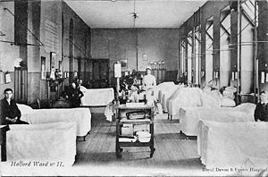
Permission was given in the same year to move the Electrical Department from the basement to the Halford Ward. On 30 June 1923, the first pioneer of the technology, Delprat Harris died at the age of 73.
Treating cancer with radium
A letter from Russell Combe, the surgeon who took the first x-ray in 1898, to the Gazette in 1923, extolled the use of x-rays in treating cancers, and that the hospital's radiologist (David Miller Muir) was in possession of a quantity of radium, which he was willing to use for such treatment. The article went on to suggest that the hospital should also acquire some radium of its own. The estimated cost of deep x-ray equipment was £1,500 and with the hospital’s own supply of radium the total cost would be £5,000. A further £5,000 was to be raised for the Electrical Department.
Throughout 1923, pressure was building for the hospital to expand its x-ray service, into the treatment of cancer, and David Miller Muir was acknowledged to be the man to do it.
Fund raising commenced in February 1924, when the Mayor Mr Philip Rowsell was recruited to the cause, with a target of £10,000. By the end of July, £3,000 had been raised, and the deep x-ray equipment had been ordered.
At a meeting in July 1923, at the inauguration of the fund, the following resolution, was moved by the Lord-Lieutenant, and was unanimously carried :
“That this meeting is of opinion that it is essential that there should be in this county a hospital equipped with everything required for the complete treatment of cancer by modern methods, and that, as a corollary, it is further essential that the County Hospital at Exeter should forthwith be provided with an adequate supply of radium, and with the necessary apparatus for deep x-ray therapy, which alone are lacking in the institution to perfect its outfit."
During August, Devon Cancer Week was held to raise funds. The Mayor said the fundraising was “for Devon, our County, to help relieve the great number of people who suffer from this dread complaint of Cancer”. Local communities throughout the county were encouraged to have a ‘flag day’ in August and September. A boxing tournament was held on the 27 August at the Civic Hall in aid of what had become the Mayor of Exeter’s Devon Cancer Fund. The silversmiths, Brufords agreed to supply a silver cup for the winner of the contest. Money was collected from across the county with many local councils donating funds, and house-to-house collections organised by the Women’s Institute.
By early 1925 the Electrical Department had completed its move to Halford Ward, and, in its own separate department, the deep x-ray equipment installed, the first in the country.
The department opens
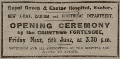
This advert appeared in the Gazette for the 3 June 1925.

The new department was opened by the Countess Fortescue to much fanfare. The new deep x-ray machine was manufactured in France by Gaiffe of Paris, and the Exeter machine was the 35th to be installed, and the first in the UK.
It is worth quoting the article from the Exeter and Plymouth Gazette, about the opening, to compare 1925 technology, with the equipment in the modern imaging department.
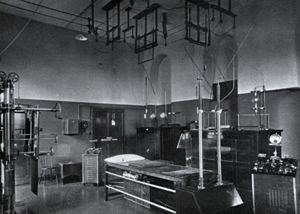
“.... machine is extremely safe, and it is impossible for a patient to be electrocuted, as none of the rays get out into the room except that required for the treatment of the patient.... The tube itself is placed in oil in a barrel-like vessel, which has lead a quarter of an inch thick, and it is impossible for a patient to do himself injury. The apparatus is worked from a small lead cubicle, thus safeguarding the operator. The department contains many other striking features, and those who remember when it was situated in the basement would hardly know it in its present home, formed from the conversion of an old double ward at an expenditure of about £1,500. It is certainly one the best departments in the country for an institution the size of the Royal Devon and Exeter Hospital, and probably the best. The electrical department includes 14 tiny platinum needles each containing 11 milligrammes of radium, the total value of £2,000. Incidentally these needles with their precious contents cost more than the deep-ray apparatus, which involved an expenditure of £1,500.
Fundraising was not finished, and in October 1925, £4,000 was still needed out of the original target of £10,000, but the department was now treating patients. Dr David Miller Muir remained at the Royal Devon and Exeter Hospital, running the x-ray imaging and cancer department until his death at the early age of 46 in 1933. Dr Charles Wroth was appointed to the post of consultant radiologist to replace Dr Miller Muir. After the wa,r the hospital was given a new machine for treating superficial cancers, from the US, under the Lend Lease Scheme. Dr Ronald Haddon was appointed as the first NHS radiotherapy consultant in 1947.
It was in the early years of the last century, that the generous donations of the people of Devon provided the first tentative steps towards treating cancer, and it is to the early pioneers, Mr Delprat Harris and Dr Miller Muir that we can thank for establishing both x-ray imaging and cancer treatment in Devon.
Sources: The Royal Devon and Exeter Hospital (1741-2006) by Andrew Knox and Christopher Gardner-Thorpe, Western Times, Exeter and Plymouth Gazette.
 The Electrical Department also housed the early x-ray equipment.
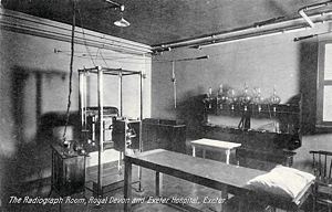
 The x-ray room was originally in the basement.
 Halford Ward before it housed the Electrical and x-ray Departments.
│ Top of Page │